


Size of this preview: 800 × 600 pixels. Other resolutions: 320 × 240 pixels | 640 × 480 pixels | 1,024 × 768 pixels.
Original file (1,024 × 768 pixels, file size: 218 KB, MIME type: image/png)
Summary
| Description |
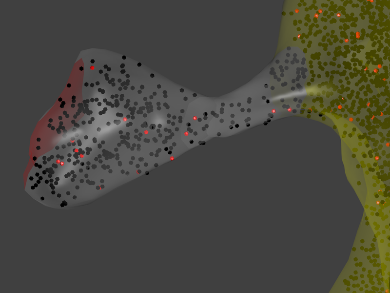
English: Screenshot from an MCell simulation of calcium signaling within the spine. Although other types of calcium-regulated molecules were included in the simulations, only CaMKII molecules are visualized. They are shown in red when bound to calmodulin and in black when unbound. The compartment is a reconstruction of a dendritic spine as presented by Kinney et al. (J Comp Neurol. 2013). The area of the postsynaptic density is shown in red, the spine head and neck in gray, and the parent dendrite in yellow. The figure was generated by visualizing the simulation results in Blender. |
| Date | |
| Source | Own work |
| Author | Mstefan |
Licensing
I, the copyright holder of this work, hereby publish it under the following license:
This file is licensed under the Creative Commons Attribution 3.0 Unported license.
- You are free:
- to share – to copy, distribute and transmit the work
- to remix – to adapt the work
- Under the following conditions:
- attribution – You must give appropriate credit, provide a link to the license, and indicate if changes were made. You may do so in any reasonable manner, but not in any way that suggests the licensor endorses you or your use.
File history
Click on a date/time to view the file as it appeared at that time.
| Date/Time | Thumbnail | Dimensions | User | Comment | |
|---|---|---|---|---|---|
| current | 22:56, 6 June 2013 | | 1,024 × 768 (218 KB) | Mstefan | User created page with UploadWizard |
File usage
The following page uses this file:
Global file usage
The following other wikis use this file:
- Usage on en.wikiversity.org
You must be logged in to post a comment.