


No higher resolution available.
EEG_fMRI.jpg (554 × 600 pixels, file size: 50 KB, MIME type: image/jpeg)
Summary
| Description |
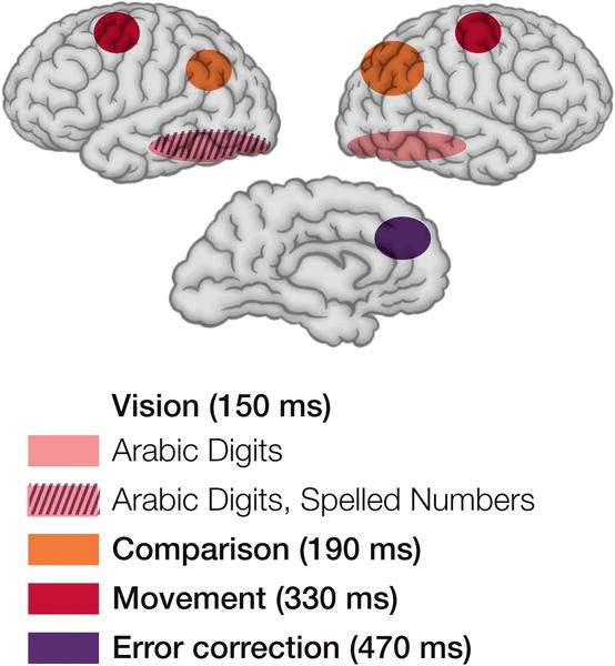
Regions of the Brain Involved in a Number Comparison Task Derived from EEG and fMRI Studies. The regions represented correspond to those showing effects of notation used for the numbers (pink and hatched), distance from the test number (orange), choice of hand (red), and errors (purple). |
|---|---|
| Source |
http://biology.plosjournals.org/perlserv/?request=get-document&doi=10.1371/journal.pbio.0030051 |
| Date | |
| Author | |
| Permission (Reusing this file) |
See below.
|
Licensing
File format
File history
Click on a date/time to view the file as it appeared at that time.
| Date/Time | Thumbnail | Dimensions | User | Comment | |
|---|---|---|---|---|---|
| current | 06:32, 2 January 2025 | | 554 × 600 (50 KB) | Jn.mdel (talk | contribs) | added notes below diagrams |
| 00:44, 17 April 2006 |  | 554 × 600 (47 KB) | Zazim (talk | contribs) | {{Information| |Description = Regions of the Brain Involved in a Number Comparison Task Derived from EEG and fMRI Studies. The regions represented correspond to those showing effects of notation used for the numbers (pink and hatched), distance from the t |
You cannot overwrite this file.
File usage
The following page uses this file: