


Size of this preview: 647 × 599 pixels. Other resolutions: 259 × 240 pixels | 518 × 480 pixels | 689 × 638 pixels.
Original file (689 × 638 pixels, file size: 148 KB, MIME type: image/png)
Summary
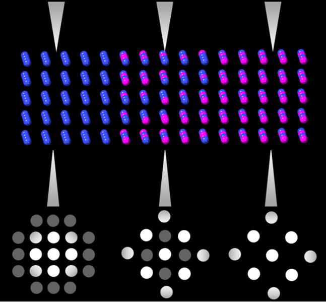
Shows the rastering of 4D scanning transmission electron microscope probe and how the diffraction pattern evolves as a function of ordering.
Licensing
I, the copyright holder of this work, hereby publish it under the following licenses:
You may select the license of your choice.
File history
Click on a date/time to view the file as it appeared at that time.
| Date/Time | Thumbnail | Dimensions | User | Comment | |
|---|---|---|---|---|---|
| current | 19:51, 13 March 2022 | | 689 × 638 (148 KB) | Matscimike (talk | contribs) | Shows the rastering of 4D scanning transmission electron microscope probe and how the diffraction pattern evolves as a function of ordering. |
You cannot overwrite this file.
File usage
The following page uses this file: