


No higher resolution available.
CT_of_lung_nodule_with_vascular_convergence_(crop).png (306 × 268 pixels, file size: 94 KB, MIME type: image/png)
Summary
| Description |
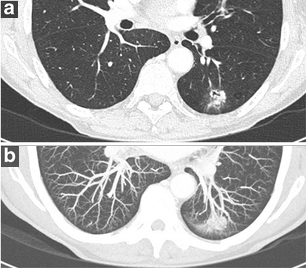
English: Original caption: Cardiac CT in 64-year-old woman with chronic cough and cardiac complaints showed a nodule in the left lower lobe. Dedicated chest CT confirmed persistence of the nodule and solitary nature. Axial CT-images in lung window setting a show a complex nodule with spiculation, pleural tags, irregular air bronchogram with bronchial interruption sign and ground glass component. Maximum intensity projection (MIP) images b better demonstrate convergence of the vessels towards the lung nodule. Malignancy was confirmed after lobectomy with histopathologic examination showing a 2.1 cm invasive adenocarcinoma. |
| Date | |
| Source |
(2017). "Evaluation of the solitary pulmonary nodule: size matters, but do not ignore the power of morphology". Insights into Imaging 9 (1): 73–86. DOI:10.1007/s13244-017-0581-2. ISSN 1869-4101.
|
| Author | Article authors: Annemie Snoeckx, Pieter Reyntiens, Damien Desbuquoit, Maarten J. Spinhoven, Paul E. Van Schil, Jan P. van Meerbeeck, Paul M. Parizel |
| Other versions |
 |
Licensing
This file is licensed under the Creative Commons Attribution 4.0 International license.
- You are free:
- to share – to copy, distribute and transmit the work
- to remix – to adapt the work
- Under the following conditions:
- attribution – You must give appropriate credit, provide a link to the license, and indicate if changes were made. You may do so in any reasonable manner, but not in any way that suggests the licensor endorses you or your use.
File history
Click on a date/time to view the file as it appeared at that time.
| Date/Time | Thumbnail | Dimensions | User | Comment | |
|---|---|---|---|---|---|
| current | 14:13, 1 May 2019 | | 306 × 268 (94 KB) | Mikael Häggström | User created page with UploadWizard |
File usage
The following page uses this file:
.png)